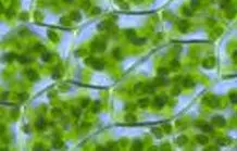
Inverted confocal microscope for high resolution fluorescence imaging. 3D image obtained from stacking of optical sections according to the confocal imaging principle, on fixed or living samples.
Associated technologies :
3D imaging
FCS
FRET
Spectral imaging
Fluorescence and contrast DIC
Its spectral detector allows in a single acquisition to detect the emission spectrum of the sample of each pixel of the image. This property allows to validate the nature of the observed compounds and facilitates the use of probes for which their analysis is based on a shift of their spectrum.
This microscope has advanced functions allowing to make live ratiometric measurements on the acquired images, length measurements, image mosaics and FRET analysis (FRET-photobleaching and FRET-sensitized emission). It is also possible to perform kinetics, record stage positions, etc.
Our confocal microscope is also equipped to perform FCS (Fluorescence Correlation Spectroscopy) analysis, allowing to define the scattering properties of single molecules.
This tool will be able to evolve towards FCCS (Fluorescence Cross-Correlation Spectroscopy) approaches for the study of protein-protein interactions.
Data sheet
Objectives :
10x Plan-Apochromat dry, N.A. 0,45 (FWD=2.1 mm)
20x Plan-Apochromat dry N.A. 0,8 (FWD=0.55 mm)
40x Plan Apo water N.A. 1,2 NA Corr FCS
63x Plan Apo oil N.A. 1,4
Illumination source full field observation at eyepieces :
- HXP100 lamp
- Observation cubes for fluorescence : DAPI, GFP, Cy3
Excitation sources confocal mode :
405 laser
Argon laser (458, 488, 514nm)
DSSP 561nm
Helium/Neon laser 633nm
Detectors :
- 2 PMT fluorescence
- 1 multi-PMT (32 GaAsP detectors) spectral detector
- 1 PMT transmission
Environnemental control : no
Acquisition type : Zeiss Zen software
- multi-modal, XYZ, spectral, time, multi-position or ratiometric confocal imaging
- 2D mosaic module
Biosafety level 1
Use & reservation
If you are interested in this microscope, please contact Christian Godon
Funding
This microscope has been funded by the Provence Alpes Côte d’Azur Region, the Bouches du Rhône General Council, the French Atomic Energy and Alternative Energies Commission, and the CNRS.

Some publications with this microscope :
- Godon et al. (2019) Under phosphate starvation conditions, Fe and Al trigger accumulation of the transcription factor STOP1 in the nucleus of Arabidopsis root cells Plant Journal, 99, 937–949 DOI : 10.1111/tpj.14374
- Kuzmic et al. (2019) Interplay between ionizing radiation effects and aging in C. elegans. Free Radical Biology and Medicine 134 (2019) 657–665. doi: 10.1016/j.freeradbiomed.2019.02.002
- Kuzmic et al. (2018) Carbonylation accumulation of the Hypsibius exemplaris anhydrobiote reveals age- associated marks. PLoS ONE 13(12): e0208617. doi: 10.1371/journal.pone.0208617
- Balzergue et al. (2017). Low phosphate activates STOP1-ALMT1 to rapidly inhibit root cell elongation. Nat Comm. 2017 May 15;8:15300. DOI: 10.1038/ncomms15300.
- Kuzmic et al. (2016) In situ visualization of carbonylation and its co-localization with proteins, lipids, DNA and RNA in Caenorhabditis elegans. Free Radical Biology and Medicine 101 (2016) 465–474. doi: 10.1016/j.freeradbiomed.2016.11.004
- Kanno et al.. (2016) A Novel Role for the Root Cap in Phosphate Uptake and Homeostasis. eLife, 5, e14577. DOI : 10.7554/eLife.14577.
Highly efficient in bright field or fluorescence (full field) imaging with image acquisition, this microscope allows to perform laser microdissection
In addition to the imaging functions (fluorescence, bright field, phase, DIC/Nomarski), this microscope has advanced functions allowing to make multiple measurements (counts, lengths, distances…) on live images.
It is also possible to perform kinetics, record stage positions, etc.
The LMD6000 also performs laser microdissection: thanks to the presence of a powerful laser, this microscope allows the cutting and sorting of sample areas into different tubes. After collection, the tissues can be used for transcriptomic, metabolomic, etc. analyses.
Associated technologies :
Laser microdissector
Multiple measurements on live images.
Technical sheet
Objectives :
1,25x HCX PL Fluotar dry, N.A. 0,04 (FWD=37 mm)
4x C PLAN dry, N.A. 0,1 (FWD=18 mm)
6,3x C PLAN dry, N.A. 0,13 (FWD=19 mm)
10x HC PL Fluotar dry, N.A. 0,3 (FWD=11 mm)
20x HCX PL FFluotar dry, N.A. 0,4 (FWD=6,9 mm)
40x HCX PL Fluotar CS dry, N.A. 0,85 (FWD=0,24mm)
63x HCX PL FluotarR L dry, N.A. 0,7 (FWD=2,6 à 1,8 mm)
63x HCX PL Apo CS water, N.A. 1,2 (FWD=0,22 mm)
Lens on request :
150x HCX PL Fluotar dry, N.A. 0,9 (FWD=0,25 mm), without coverslip
Illumination source full field observation at the eyepieces :
- HXP100 lamp
- Observation cubes for fluorescence: DAPI, GFP, DsRed, on request: other filter cubes possible
- cubes for microdissection: DAPI, GFP, CY3, Alexa Fluor
Camera : Leica DFC7000T
Environnemental control : no
Acquisition type : Leica LAS software
Use & reservation
If you are interested in this microscope, please contact Christian Godon
Funding

This microscope is attached to the Heliobiotec* platform and has received funding from the European Union (European Regional Development Fund), the Provence Alpes Côte d’Azur Region, the French Ministry of Research, and the French Atomic Energy and Alternative Energies Commission.
Some publications with this microscope
- Kanno et al.. (2016) A Novel Role for the Root Cap in Phosphate Uptake and Homeostasis. eLife, 5, e14577. DOI : 10.7554/eLife.14577.
- Hirsch et al. (2011) A Novel fry1 Allele Reveals the Existence of a Mutant Phenotype Unrelated to 5’>3’ Exoribonuclease (XRN) Activities in Arabidopsis thaliana Roots. PLoS ONE 6(2): e16724. doi:10.1371/journal.pone.0016724
Dedicated to luminescence approaches with tissue/cellular resolution, this microscope allows to obtain very good quality images even for samples with low luminescence.
It also allows to obtain bright field and transmission fluorescence (GFP) images.
A temperature / CO2 control chamber can be installed on request. It allows to maintain cell cultures or tissue sections in a controlled environment throughout the experiment. The LV200 housing perfectly protects the sample and the optics from external light.
The optical path from the object to the CCD camera is straight, optimized to the shortest possible path to ensure the least amount of photon loss.
The excitation and emission filter holder for GFP allows fluorescence image acquisition in addition to luminescence image acquisition. Transmitted light image acquisition (brightfield illumination with phase contrast rings) allows to produce superpositions with images obtained in luminescence and fluorescence.
Technical data sheet
Objectives:
4x UPLSAPO dry, N.A. 0,16 (FWD=13 mm)
20x LUCPLFLN dry N.A. 0,45 (FWD=6,6-7,8 mm)
40x LUCPLFLN dry N.A. 0,6 (FWD=2,7-4 mm)
Lens on request :
10x UPLFLN dry N.A. 0,3 (FWD=10 mm)
40x UPlanApo oil N.A. 1
60x UPLAPO dry, N.A. 0,9 (FWD=0,13 mm)
60x UPLAPO oil, N.A. 1,4 (FWD=0,13 mm)
100x UPLAPO oil, N.A. 1,35 (FWD=0,13 mm)
Source of illumination :
- HXP100 lampe
- observation cubes for fluorescence: GFP
Camera : ANDOR iKon-M DU934
Environmental control : yes
Use & reservation
If you are interested in this microscope, please contact Hélène Javot
Funding

This microscope was funded by the Commissariat à l’énergie atomique et aux énergies alternatives, and the CNRS (Interface physique-chimie-biologie program).
Some publications with this microscope
- Kanno et al.. (2016) A Novel Role for the Root Cap in Phosphate Uptake and Homeostasis. eLife, 5, e14577. DOI : 10.7554/eLife.14577.
- Hirsch et al. (2011) A Novel fry1 Allele Reveals the Existence of a Mutant Phenotype Unrelated to 5’>3’ Exoribonuclease (XRN) Activities in Arabidopsis thaliana Roots. PLoS ONE 6(2): e16724. doi:10.1371/journal.pone.0016724
This inverted microscope allows the observation of fluorescent samples (or not) under slide/lamellar. Apotome function to make optical sections and 3D images.
Associated technologies :
Apotome
3D imaging
Fluorescence and DIC contrast
This inverted microscope allows the observation of fluorescent samples (or not) under slide/lamella. The apotome function allows to generate optical sections eliminating the background noise of the image (blur). The apotome principle, although very different from a confocal microscope, allows to obtain high quality images.
Transmission illumination, phase contrast and DIC
The image acquisition software has advanced functions such as: automation of acquisition sequences, custom programs, image mosaics, series of optical sections…
Data sheet
Objectives :
10x Ph1 M27 EC Plan Neofluar dry, N.A. 0,3 (FWD=5,2mm)
20x M27 Plan Apochromat dry, N.A. 0,8 (FWD=0,55mm)
40x M27 EC Plan Neofluar dry, N.A. (FWD=0,71mm)
63x DIC M27 Plan Apochromat oil, N.A. 1,40 (FWD=0,19mm)
100x M27 Plan Apochromat oil, N.A. 1,40 (FWD=0,17mm)
Illumination source full field observation at the eyepieces :
- HXP100 lamp
- Observation cubes for fluorescence : Dapi-GFP-CY3-CY5
Camera :
Environnemental control : no
Acquisition type : Zeiss Zen software
- 2D mosaic module
Use & reservation
If you are interested in this ultramicrotome and/or resin fixation protocols, please contact Olivier Simon
Funding

This microscope was financed by the IRSN.
Macroscopic fluorescence imaging
This macroscope allows the observation of fluorescent samples (or not) at a macroscopic level (from a few cells to whole organisms). Very good quality images can be obtained from samples under coverslip, but also through Petri dishes, or from plants in pots.
The apotome function (based on structured illumination) allows to generate optical slices eliminating the background noise of the image (blur). The apotome principle, although very different from a confocal microscope, allows to obtain high quality images.
The image acquisition software has advanced functions such as: automation of acquisition sequences, custom programs, image mosaics, series of optical sections…
Transmission illumination, pseudo dark field and contrast method
Fluorescence with fibered source for DAPI-GFP-RFP
Monochrome camera, with color reconstruction by 3 images (R+G+B)
Color camera with or without fluorescence imaging
0.5x objective for final magnification from 3.5x to 56x and 2.3x objective for final magnification from 16x to 258x
Apotome function usable only with the 2.3x objective, in fluorescence mode.
Associated technologies :
Wide field image, mosaic
3D imaging
Fluorescence
Apotome (structured illumination)
Color photo camera Sony
Technical data
Lenses :
0.5x Plan-Apo lens, N.A. 0.125 (FWD 11.4 mm)..
Lens 2.3x PlanNeoFluar, N.A. 0.57 (FWD 10.6 mm).
Cameras :
- Zeiss monochrome camera
- Sony 7 III color camera : 24,2 MP, Iso 100-51200 and 4K/UHD video.
Illumination/Excitation Source :
HXP 200 lamp
Observation cubes for fluorescence : DAPI, GFP, mRFP and GFP LP
Apotome module
Motorized stage
Acquisition type :
- Zeiss camera : Zeiss Zen software 2D mosaic and 3D apotome module
- Sony camera : Sony software
Use & reservation
If you are interested in this microscope, please contact Serge Chiarenza
Some linked publications
- Hanchi et al. (2018). The phosphate fast-responsive genes PECP1 and PPsPase1 affect phosphocholine and phosphoethanolamine content. Plant Physiology. 176(4):2943-2962. DOI: 10.1104/pp.17.01246.
- Balzergue et al. (2017). Low phosphate activates STOP1-ALMT1 to rapidly inhibit root cell elongation. Nat Comm. 2017 May 15;8:15300. DOI: 10.1038/ncomms15300.
- Kanno et al.. (2016) A Novel Role for the Root Cap in Phosphate Uptake and Homeostasis. eLife, 5, e14577. DOI : 10.7554/eLife.14577.