
Soil bacteria interact with metals and radionuclides (RN) and can modify their chemical speciation, thus modulating their mobility and bioavailability. Radionuclides are chemo- and radio-toxic elements that can in turn induce cellular stress and against which certain species exhibit a high tolerance capacity. At the environmental level, the role of bacteria in the transfer of RN, particularly in the soil and to plants, is still very poorly understood. At the cellular level, the molecular mechanisms involved in detection, transport, response and tolerance to RN are not yet characterized.
In this context, we are studying :
- The cellular response and mechanisms of tolerance to uranium in strains isolated from radionuclide-rich soils using a multidisciplinary approach combining molecular biology, biochemistry, genomics, proteomics and biophysics (Gallois et al. J Proteomics 2018)
- The mechanisms of sequestration of alkaline earths (in particular calcium and strontium) by a photosynthetic bacterium and its potential for bioremediation (Mehta et al. 2019, HARLEY project)
- The role of bacteria in the transfer of uranium to plants (RADONORM and INSPECT projects)
For the experimental part, we are equipped with a laboratory dedicated to the handling of bacteria in the presence of radioactive elements (SALTO).
1- COBALT OXIDE PARTICLES TOXICITY – EDF FUNDING
The existence of cases of human contamination by accidental inhalation of cobalt in the form of radioactive oxides in EDF installations justifies the need for research on the toxicity and behaviour of these particles. We previously addressed the chemical toxicity of cobalt oxides on lung cells using the stable cobalt isotope 59Co. Even if the effects of the emitter 60Co have been widely described in the literature for external irradiation, these effects are not known after internal contamination.
Internal contamination by 60Co particles results in chronic, localized irradiation due to the long-term retention of the particles in the body. Therefore, the in vivo behaviour of 60Co particles and the design of specific decorporants represent a strong interest both for nuclear manufacturers such as EDF and for public health. The objective of our project is to identify and test in vitro and in vivo molecules that increase the dissolution of Co particles to decrease pulmonary retention and promote the excretion of cobalt. Through an extensive screening based on dissolution studies of other metal oxides, we selected molecules with different properties, such as redox potential and chelating ability, and greatly improved the dissolution of the particles as compared to controls (1). The ongoing work is now focused on dissolution in cellular models, and on decorporation studies in vivo to identify new therapeutic strategies.

2- TRITIATED DUST AND DISMANTLING
Launched in September 2017, the European Horizon 2020 TRANSAT (TRANSversal Actions for Tritium) project will last 48 months with a total budget of nearly €4 million. Coordinated by CEA, it gathers 18 partners from 8 European countries.
During the decommissioning of nuclear facilities, operations are intended to remove or eliminate any tritiated material. These operations generate fine airborne dust, namely aerosols. The workpackage 3 is dedicated to the study of the consequence of a release of tritiated stainless steel and cement particles in terms of radiotoxicology and ecotoxicology (2). The outcomes foreseen in this project will help radiation protection authorities, IAEA and other nuclear safety advisory organisms to assess more precisely the radiobiology, dosimetry, genotoxicology and ecotoxicology of tritiated micron and sub-microns particles.
We are involved in this project at two levels :
1/ Leading of workpackage
2/ Study of toxicity, genotoxicity and behaviour of tritiated particles in human lung models (in collaboration with AMU IMBE)
– Use of two in vitro models: the immortalized BEAS-2B cell line and the 3D in vitro cell model of the human airway epithelium MucilAir® to study the contribution of the chemical and the radiological stress by the use of tritiated and non tritiated particles
– Assessment of short and long terms effects and reversibility of the toxic effects after an acute exposure
– Evaluation of the tritium transfer through the lung epithelial barrier in a kinetic mode
– Definition of the mode of cytotoxic effects using a combination of assays including cytotoxic (metabolism, epithelial integrity), clonogenic (cloning efficiency) and oxidative stress related (ex: GSH/GSSG ratio) assays.
– Assessment of genotoxic hazards by standard genetic toxicology methods (comet and micronucleus assays using centromeric probes/ anti-kinetochore staining), measurements for epigenetic modifications (DNA methylation, chromatin profiling, microRNA content) to address DNA regulation, primary DNA lesions and chromosome damage

BIBLIOGRAPHY :
- Van Der Meeren A, Lemaire D, Coudert S, Drouet G, Benameur M, Gouzerh C, et al. In vitro assessment of cobalt oxide particle dissolution in simulated lung fluids for identification of new decorporating agents. Toxicol In Vitro. 2020;66:104863.
- Liger K, Grisolia C, Cristescu I, Moreno C, Malard V, Coombs D, et al. Overview of the TRANSAT (TRANSversal Actions for Tritium) project. Fusion Engineering and Design. 2018.

Radionuclides and certain metal cations present in the environment, sometimes on a large scale, pose toxicity problems. In the team, we study the molecular determinants of the interaction between metal cations and proteins in order to better understand the mechanisms of toxicity, notably by competition with biological metal cations, or with the aim of developing affine and selective proteins for the biodetection or bioremediation of toxic metals.
These studies involve combined approaches of protein engineering and characterization by different methods, UV-Vis spectroscopy, Fluorescence, calorimetry, ESI-MS, FTIR.
The projects in progress concern :
- The interaction properties between variants of the EF-hand motif that normally fixes calcium and uranium (collaboration with LILA, DEN-Marcoule) as well as Ca / Sr selectivity (DEMETERRES project).
- The interaction properties of plutonium and proteins2 (coll. LILA, DEN-Marcoule).
- The properties of interaction with alkaline of a protein that could be key in the precipitation of these compounds as carbonates in cyanobacteria (ANR Harley, K. Benzerara IMPMC)
- The role of periplasmic prtein in the incorporation of lanthanides in P. putida (EC2CO project Lanthanomics, P. Billard Univ Nancy).
- Phage-assisted directed evolution to modify the metal selectivity of proteins involved in the response to metal stress (ANR project BioBrickevolver, P. Nicolas, MaIAGE, INRAE). These proteins could serve as a basis for developing cobalt biosensors.
1 Sauge-Merle et al. Chem. Eur. J. 2017, 23, 15505 – 15517
2 Sauge-Merle et al. Dalton Trans. 2017, 46, 1389-1396
At the IPM, a first uranyl fluorescent biosensor was obtained, based on the engineering of uranyl affine and selective sites1,2,3. This biosensor, inspired by the calcium fluorescent biosensor of Tsien’s team (Nobel Prize in Chemistry in 2008)4, is constructed on the basis of the modified N-terminal domain of calmodulin framed by two fluorescent proteins (GFPs, Figure 1A). The metal binding leads to a fluorescence transfer called FRET (Fluorescence Resonance Energy Transfer) between the 2 GFPs, which gives access to the amount of metal bound (Figure 1B). The major assets of this probe are a fast and ratiometric response, implying a result independent of the quantity of sensor.

Figure 1. Fluorescent uranyl biosensor
A. Functioning of biosensor. B. Fluorescence spectra measured int the presence of Ca2+ or UO22+
Its in vitro characterization showed an apparent affinity for uranyl in the micromolar range, a very marked selectivity for uranyl with respect to other cations tested (Ca2+, Mg2+, K+, Na+) and a good robustness with a working pH range between 5 and 7.5.
This biosensor was then validated in vivo for the detection of uranyl in the roots of the model plant Arabidopsis thaliana (Thesis R. Cherif with paper in preparation, collaboration H. Javot (SAVE/BIAM)). Confocal microscopic analysis of the fluorescence of roots expressing the biosensor shows that exposure of roots to 10 µM uranyl for 24H leads to a significant increase in FRET in the presence of uranyl. This result indicates that the biosensor is able to measure a flow of uranyl from the culture medium to the root (Figure 2), while the fraction of uranium incorporated in the root under these conditions is estimated at 1%. Kinetic and microfluidic dose-response studies are under way to specify this uranyl flux.

Figure 2. Effect of exposing plant roots to a 10 µM uranyl solution for 24 hours
A. Roots observed under the confocal microscope. B. Statistical analyses of image processing by Image J and R
A new project begins in the framework of Nicolas Jans’ thesis and aims to express the fluorescent uranyl biosensor in zebrafish larvae (Danio rerio) for the on-line detection of bioavailable uranium (collaborations DAM / TEFOR Paris Saclay / IRSN – thesis FOCUSDEM, ECCOREV2020).

1. Pardoux R, Sauge-Merle S, Lemaire D, Delangle P, Guilloreau L, Adriano JM, Berthomieu C (2012) PLoS ONE, 7(8) e41922
2. Pardoux R, Sauge-Merle S, Lemaire D, Berthomieu C, Guilbaud P, Delangle P, Bremond N, Beccia, M-R (2014) Brevet européen EP2784094A1 + WO2014155356 A8 (extension internationale)
3. Sauge-Merle S, Brulfert F, Pardoux R, Solar PL, Lemaire D, Safi S, Guilbaud P, Simoni E, Merroun ML, Berthomieu C (2017) Chemistry, 23, 15505-15517
4. Miyawaki A, Llopis J, Heim R, McCaffery JM, Adams JA, Ikura M, Tsien RY (1997) Nature, 388, 882-887
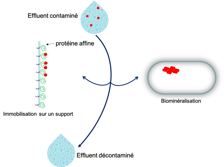
Different projects developed in the team, ranging from the study of environmental bacteria to protein engineering and synthetic biology, could feed in the more or less long term bioremediation applications for the treatment of water or effluents contaminated with metals and NRs.
These projects concern :
- The sequestration of NRs by bacteria capable of trapping them by biomineralization. These aspects are developed on different bacterial models for uranium and 90Sr in particular.
- The identification and characterization of uranium affine proteins synthesized by environmental bacteria in response to uranyl stress.
- The engineering of chelating architectures allowing the selective fixation of a metal or RN of interest.

Several of the team’s themes are supported by the study of structure-property relationships of proteins involved in the response to metals. Biochemical approaches are combined with biophysical approaches such as fluorimetry, denaturing or native mode mass spectrometry preserving protein-cofactor interactions or Fourier transform infrared spectroscopy.
These biophysical approaches are complementary to structural approaches such as X-ray diffraction, to study the properties of active sites or enzymatic mechanisms. They are dedicated to the study of metalloproteins or photoenzymes such as photosystems or the fatty acid photodecarboxylase (FAP) in collaboration with different teams of the BIAM.
Identification and exploitation of biological systems of acquisition and complexation of Rare Earth Elements (RE) by bacteria for selective extraction in the context of recycling processes of FeNdB magnets.
Loïc Aleman – 2021 2025
Development of fluorescent uranyl biosensor for environmental monitoring.
Nicolas Jans-Robinson – 2021 2025
Towards a breakthrough effluent treatment process : use of cyanobacteria selectively precipating strontium versus calcium.
Edern Pamart – 2024
Plutonium speciation in complex media.
Loïc Daronnat – 2020 2023
Characterization of UipR, S, A proteins : a system involved in uranium tolerance and iron homeostasis in Microbacterium.
Abbas Mohamad Ali – 2023
http://www.theses.fr/2023AIXM0228
Study of structure-function relationship in photosynthetic proteins by differential spectroscopy in the medium and low frequency IR range.
Lidia Zuccarello – 2016 2019
http://www.theses.fr/2019AIXM0107
Cellular response of environmental Microbacterium isolates to uranium exposure.
Nicolas Gallois – 2015 2018
http://www.theses.fr/2019AIXM0107
Analysis of the bacterial biodiversity of a contaminated soil in the Chernobyl area and characterization of the interaction of a strain of Microbacterium with uranium.
Nicolas Théodorakopoulos – 2010 2013
http://www.theses.fr/2013AIXM4108
Optimization of a uranium chelating protein site derived from calmodulin.
Romain Pardoux – 2009 2012
http://www.theses.fr/2012AIXM4116
Analysis and exploitation of bacterial populations in uranium-rich soils: selection of a model species.
Laure Mondani – 2007 2010
http://www.theses.fr/2010AIX22113
Study of protein-metal interactions by infrared spectroscopy in the low frequency range.
Laure Marboutin – 2003 2006